Muscles of Facial Expression | Facial Anatomy for Massage Professionals
Sep 26, 2026
Understanding facial muscles anatomy changes the way a professional approaches facial massage.
Instead of memorizing a sequence of movements, an esthetician or massage therapist can begin to understand what tissue is under the hands, what movement that muscle creates, where it attaches, and how neighboring muscles work together.
For facial massage professionals, the muscles of facial expression are especially important because many of them have an unusual relationship with the skin.
“Because the muscles insert in the skin rather than on bone, when they contract, the skin moves to create facial expression.”
OpenStax: Muscles That Create Facial Expression
That relationship between muscle, connective tissue, and skin is one reason facial anatomy for estheticians and facial anatomy for massage therapists deserves more attention than simply learning the names of the muscles.
What Are the Muscles of Facial Expression?
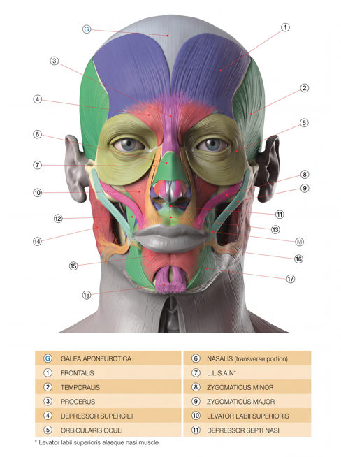
The face contains multiple layers of skeletal muscles responsible for facial movement, communication and expression.
The National Library of Medicine's StatPearls review describes facial muscles as muscles that connect facial skin with the skull and perform functions including expression and mastication. It broadly divides them into muscles of facial expression and muscles of mastication.
NCBI Bookshelf: Anatomy, Head and Neck — Facial Muscles
Most muscles of facial expression are innervated by the facial nerve, cranial nerve VII. The muscles of mastication, including the masseter and temporalis, are primarily innervated through the mandibular division of the trigeminal nerve, cranial nerve V.
This distinction becomes particularly useful when learning facial massage because the muscles we palpate around the forehead, eyes, cheeks, mouth and jaw do not all perform the same type of work.
Why Are Facial Muscles Different From Many Other Skeletal Muscles?
Many skeletal muscles move a joint by attaching bone to bone.
The muscles of facial expression are different.
TeachMeAnatomy explains that these muscles are located largely in the subcutaneous tissues of the face and typically “originate from bone or fascia and insert onto the skin.”
TeachMeAnatomy: The Muscles of Facial Expression
When these muscles contract, they move the skin and surrounding soft tissue. This allows us to smile, squint, raise the eyebrows, purse the lips and produce countless subtle facial expressions.
For a facial massage professional, that anatomy matters. We are not working with isolated muscles arranged like separate straps. We are working with an interconnected system of muscles, fascia, skin and other soft tissues.
Important Facial Muscles for Massage Professionals
Frontalis
The frontal belly of the occipitofrontalis raises the eyebrows and contributes to horizontal movement and wrinkling of the forehead.
For professionals studying forehead massage, it is useful to understand not only where the frontalis lies but also its relationship with the scalp through the epicranial aponeurosis.
Corrugator Supercilii
The corrugator supercilii draws the eyebrows medially and downward.
It is one of the small muscles involved in movement around the glabella and eyebrow region and is highly relevant when studying the anatomy behind repeated frowning expressions.
Orbicularis Oculi
The orbicularis oculi surrounds the eye and helps close the eyelids.
Because the periorbital area contains delicate tissues and important anatomical structures, knowing the position and function of this muscle is particularly important when working around the eyes.
Zygomaticus Major and Minor
The zygomatic muscles help elevate structures around the upper lip and corner of the mouth and contribute to smiling expressions.
They run across the cheek region and interact functionally with several other muscles around the mouth.
Orbicularis Oris
The orbicularis oris surrounds the mouth and participates in movement and control of the lips.
Unlike a simple circular band, this region contains complex interconnections between multiple facial muscles.
Buccinator
The buccinator muscle forms an important muscular component of the cheek.
It compresses the cheek toward the teeth and assists with functions such as chewing, blowing and controlling material within the oral cavity. OpenStax notes that the buccinator “compresses the cheek” and contributes to chewing.
Its location makes the buccinator particularly important in the study of buccal massage anatomy and intraoral techniques.
Mentalis
The mentalis is located in the chin region. It helps elevate and protrude the lower lip while moving the skin of the chin.
Understanding its position is useful when examining tension and movement patterns around the lower lip and chin.
Platysma
The platysma is a broad, thin superficial muscle extending from the upper chest and neck toward the lower face and mandible.
Its anatomical connection between the neck and lower face is one reason facial massage education should not treat the face and neck as completely separate regions.
What About the Masseter and Temporalis?
The masseter and temporalis are not primarily classified as muscles of facial expression. They are muscles of mastication.
The masseter contributes strongly to closing the jaw, while the temporalis also participates in mandibular movement. Cleveland Clinic identifies the masseter, temporalis and pterygoid muscles among the major muscles used for chewing.
Cleveland Clinic: Facial Muscles — Anatomy and Function
For facial massage professionals, however, the distinction between expression muscles and mastication muscles does not mean these structures should be studied separately.
Jaw movement, facial expression, the cheek, lips and neck all operate within the same anatomical region.
Understanding those relationships is particularly important for professionals working with facial sculpting, jaw tension or intraoral massage.
Why Facial Anatomy Matters for Estheticians
Facial anatomy for estheticians should go beyond identifying muscles on an illustration.
Understanding muscle location and function can help an esthetician better understand what they are palpating and why different regions of the face move the way they do.
It can also help professionals distinguish between structures that may look similar on a simplified anatomy chart but differ greatly in depth, function and surrounding anatomy.
This does not mean that every facial treatment needs to become a clinical anatomy lesson. It means that technique becomes more intentional when the practitioner understands the tissues being worked with.
Why Facial Anatomy Matters for Massage Therapists
The same principle applies to facial anatomy for massage therapists.
Massage therapists already work with muscular anatomy throughout the body. Applying those same anatomical principles to the face requires recognizing that facial muscles are relatively small, closely interconnected and often attached partly to skin or neighboring soft tissues rather than functioning like the larger muscles of the limbs.
A practitioner who understands facial muscles anatomy can make better-informed decisions about positioning, direction of movement, pressure and how external facial work relates to the jaw, scalp and neck.
Learn the Anatomy Before Learning the Sequence
A facial massage protocol can show you where to put your hands.
Anatomy helps you understand why your hands are there.
That difference becomes increasingly important as facial massage techniques become more specialized.
For professionals learning sculptural facial massage, buccal massage or intraoral work, understanding the muscles of facial expression, muscles of mastication and surrounding soft-tissue anatomy provides the foundation for safer and more intentional practice.
At The Sculpt Lab Academy, facial massage education begins with anatomy for exactly this reason. Techniques become easier to understand, adapt and apply when the practitioner can visualize the structures underneath their hands.
This article is intended for professional education and general anatomical information. It does not replace medical education, diagnosis, or individualized medical advice.